OCT is a 3D ultrasound scan of your retina, in particular your optic nerve and your macula.
Conventional methods of eye examination available used in most opticians, including retinal photography, only able the optometrist to see what is happening on the surface of your retina, when a lot of conditions of the eye start at a much deeper level.
OCT is a safe and non-invasive method of precisely analysing the microscopic layers of your retina. Using this amazing piece of advanced technology enables us to diagnose so many more eye problems at the earliest possible stage and in some cases up to 5 years earlier than would be possible otherwise.
The earlier any eye condition is detected, the earlier your treatment commences, thus having a much greater chance of a successful outcome. In many cases, early detection can even prevent a condition from developing at all, thus helping us to do our job, which is to help maintain your sight.
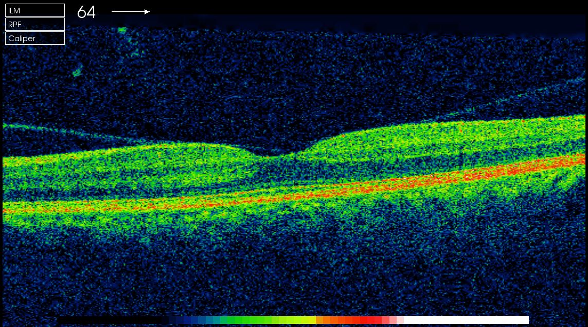
Image showing vitreomacular adhesion which could lead to vitreomacular traction.
Common conditions identified through regular OCT screening include:
1. Age-related macular degeneration
Macular degeneration causes the gradual breakdown of the macular (the central portion of the eye). OCT can identify this condition and its type (there are two types, wet and dry) and also monitor its progress, for example if you are undergoing treatment for such a condition. Unfortunately the risk of developing macular degeneration increases with age, and it is the most common cause of vision loss in individuals over the age of fifty.
We believe that OCT screening is the earliest way to detect any macular changes such as macular degeneration.
2. Glaucoma
Glaucoma damages the optic nerve at the point where it leaves the eye. Recent statistics suggest that some form of glaucoma affects around two in every 100 people over the age of 40. The danger with chronic glaucoma is that there is no pain and your eyesight will seem to be unchanged, but your vision is being damaged. An OCT examination will confirm if you are at risk, or indeed what stage of glaucoma you may have.
OCT examination can in cases detect glaucoma up to 50% earlier than conventional methods. In most cases this is a condition which is relatively easy to treat. OCT screening has the potential to give you a 50% larger window to find the right course of treatment for you, thus making the condition easy to manage.
3. Diabetes
Diabetic retinopathy is a major cause of visual impairment among adults. Here in the UK, more than two million people have been identified as having diabetes. OCT examination enables early detection, which greatly improves the success rate of treatment.
4. Macular holes
A macular hole is a small hole in the macular – the part of the retina which is responsible for our sharp, detailed, central vision. This is the vision we use when we are looking directly at things, when reading, sewing or using a computer. There are many causes of macular holes. One is caused by vitreous detachment, when the vitreous pulls away from the back of the eye and sometimes it does not ‘let go’ and eventually tears the retina, leaving a hole. Extreme exposure to sunlight (for example staring at the sun during an eclipse) can also cause a macular hole to develop.
5. Vitreous detachments
Vitreo-macular traction can clearly be diagnosed through OCT providing invaluable information about the current relationship between the vitreous and the retinal surface of the eye. As people get older the vitreous jelly that takes up the space in our eyeball can change. It becomes less firm and can move away from the back of the eye towards the centre, in some cases parts do not detach and cause ‘pulling’ of the retinal surface. The danger of a vitreous detachment is that there is no pain and your eyesight will seem unchanged but the back of your eye may be being damaged.
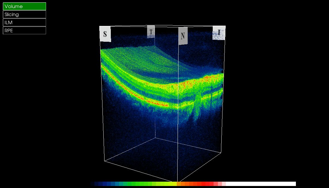
Image showing a 3D cross section of the macula
The retina is the light-sensitive tissue that lines the inside of the eye. The retina functions in a manner similar to film in a camera. The optical elements within the eye focus an image onto the retina of the eye, initiating a series of chemical and electrical events within the retina. Nerve fibers within the retina send electrical signals to the brain, which then interprets these signals as visual images.